Microscopy and Imaging Facility
From high-resolution live-cell fluorescence imaging to routine brightfield and dissecting microscopy, our light microscopy provision spans two buildings and a broad range of research and teaching needs.
What you can do here
Our microscopy provision covers the full range of routine and specialist light microscopy needs, from everyday sample observation through to high-resolution multi-channel fluorescence imaging and single-molecule manipulation.
Our microscopes
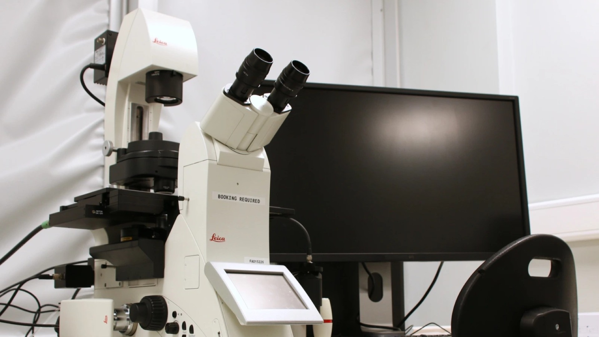
Our two Leica DMi8 systems support high-resolution fluorescence and live-cell imaging. Each is fitted with monochrome and colour cameras, and a motorised stage and objective turret for precise, repeatable positioning.
One system uses a Sutter Lambda DG-4 high-speed wavelength switching lamp for rapid multi-channel imaging; the other uses a Leica EL6000 external fluorescence light source.
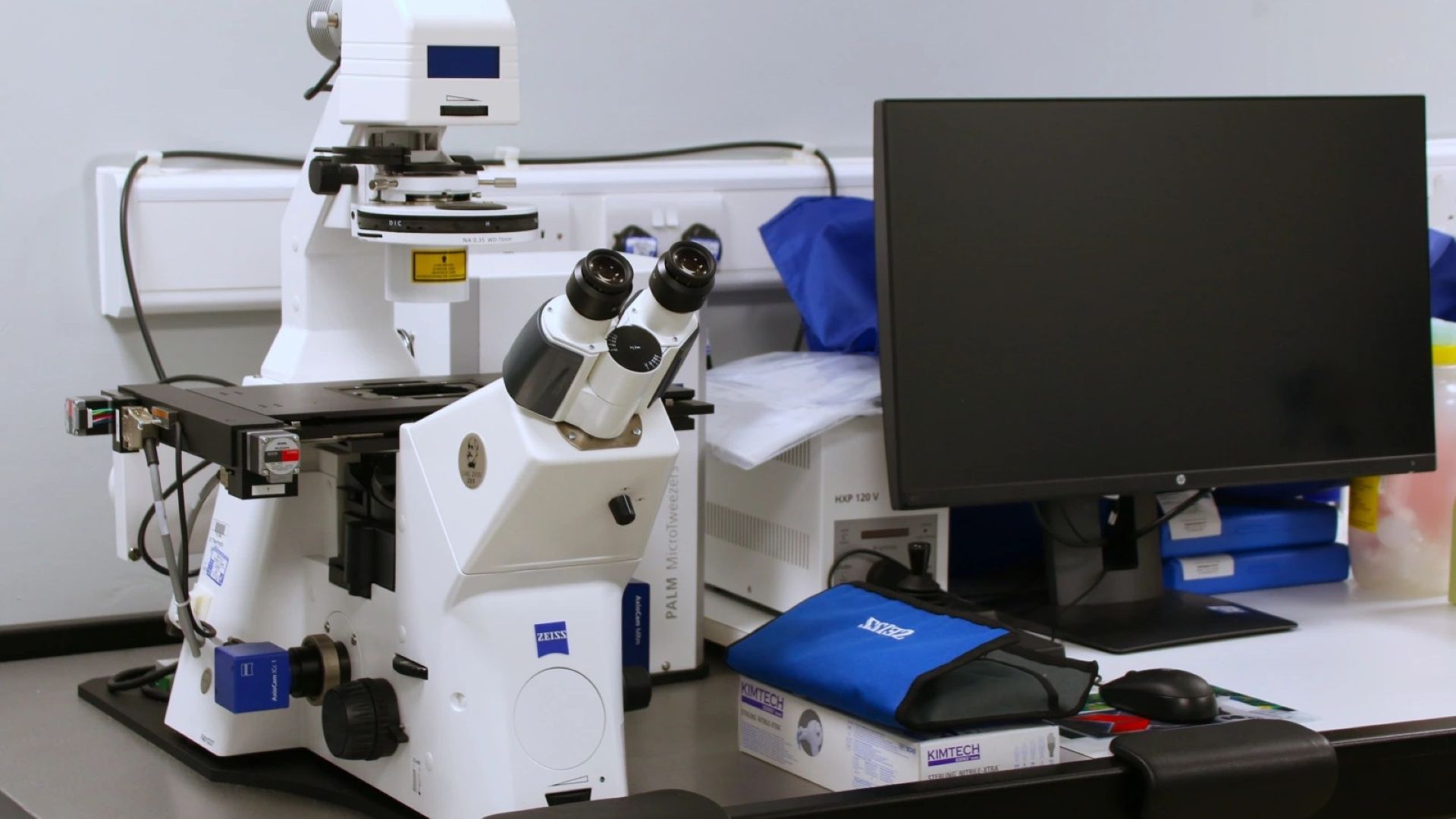
Built around a Zeiss Axio Observer inverted microscope with monochrome fluorescence imaging, the PALM MicroTweezers uses a focused infrared trapping laser to capture and manipulate microscopic particles in suspension, without physical contact. Suitable for cells, bacteria, and other sub-cellular particles, the system allows precise positioning and movement of your sample using the force of light alone and can run alongside fluorescence imaging for simultaneous visualisation.
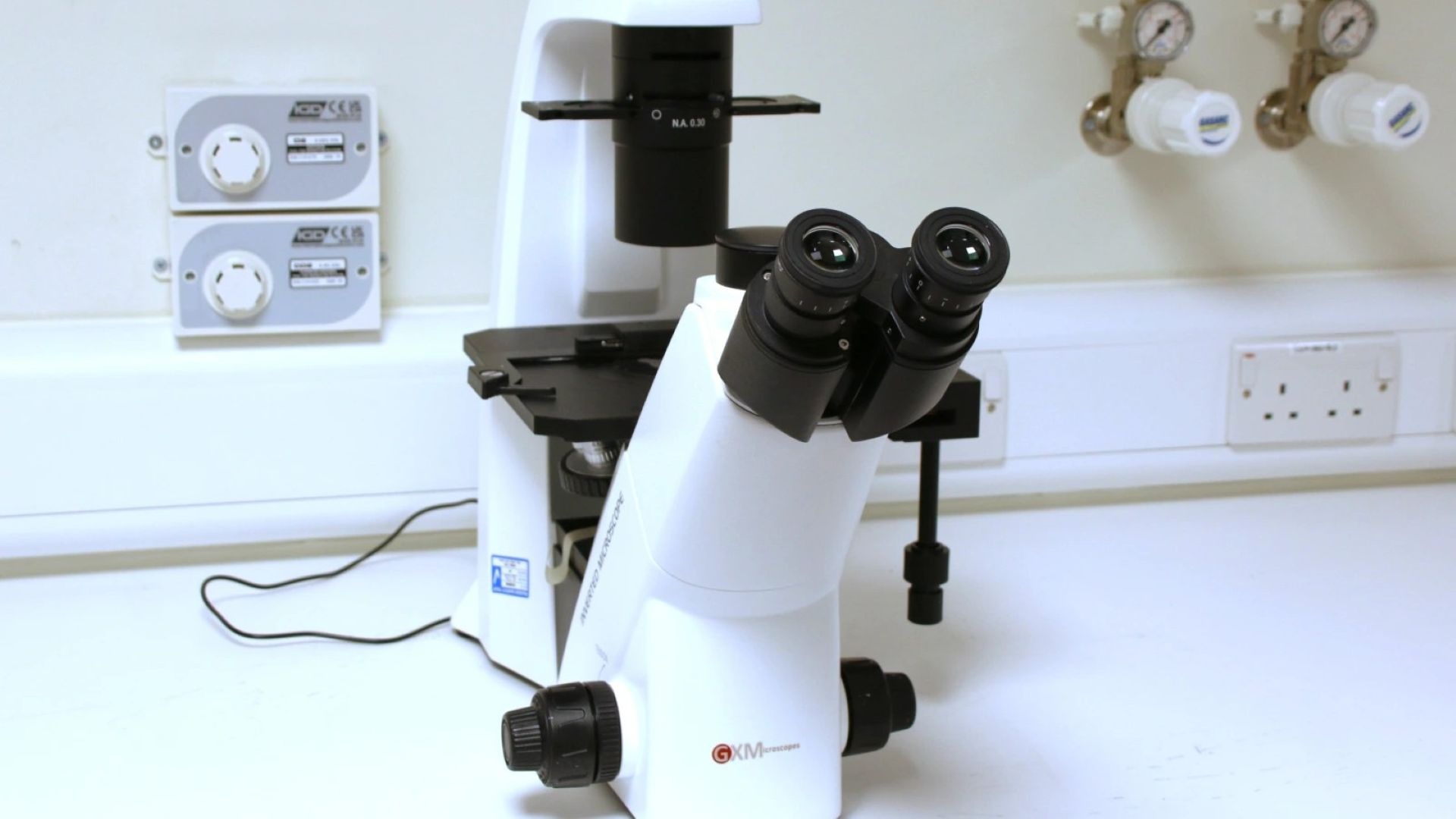
A research-grade inverted microscope with a compact, low-footprint design well suited to tissue culture rooms. Fitted with infinity-corrected plan achromatic objectives (4×, 10×, 20×, 40× — including phase contrast), a mechanical stage with culture dish holders, and a trinocular head with camera port. 5W LED illumination. Suitable for routine cell culture monitoring and research-level brightfield and phase contrast work.
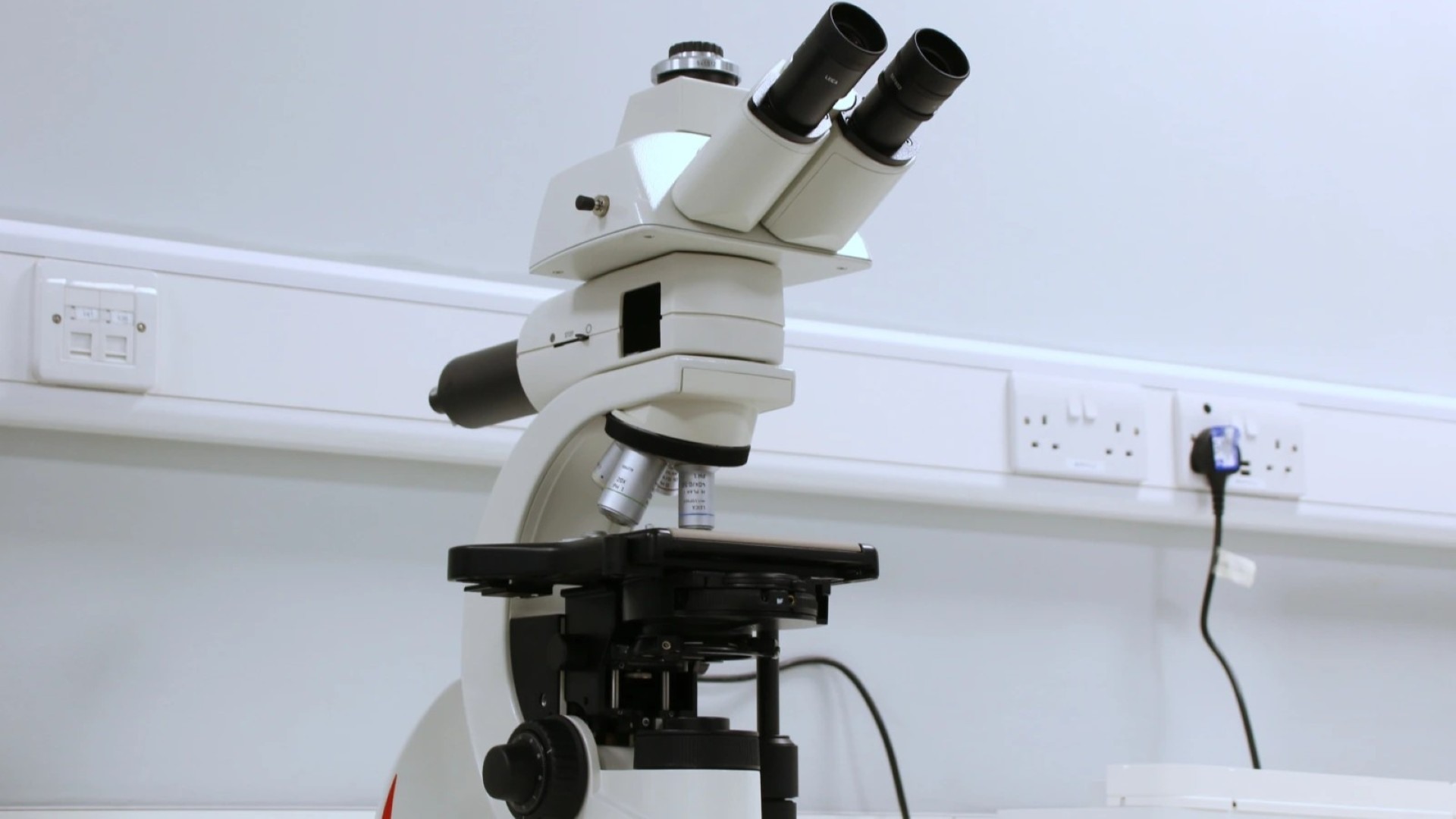
An ergonomic upright compound microscope with long-life LED illumination providing consistent colour temperature and near-daylight brightness. Height-adjustable focus knobs and customisable ergonomic configuration make it well suited to extended use sessions. Supports a range of contrast techniques and objectives. Certified for in-vitro diagnostics applications including cytology, haematology, and pathology.
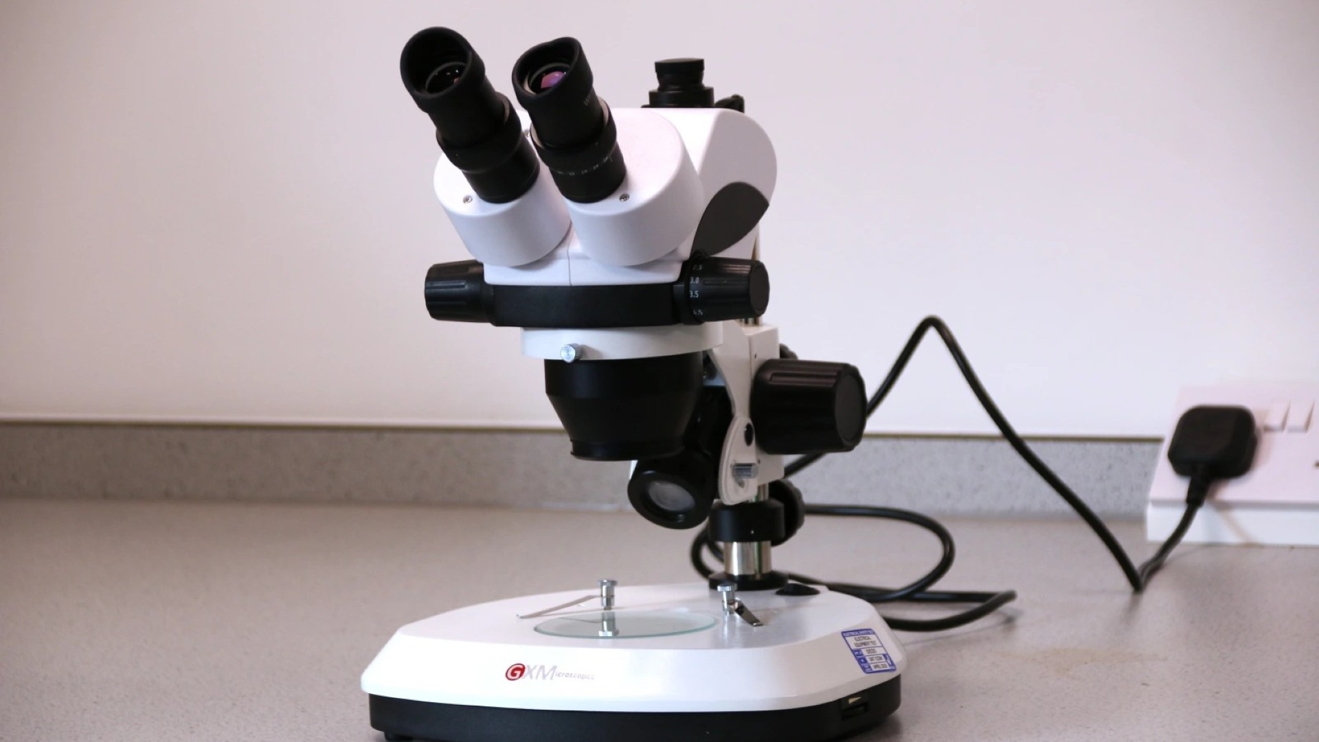
A stereo zoom dissecting microscope with a continuous magnification range of 7×–45× (standard 10× widefield eyepieces), controlled by a smooth zoom knob for rapid magnification changes without swapping objectives. Built-in incident and transmitted halogen illumination makes it versatile for a wide range of biological and materials specimens. The 88mm working distance provides comfortable space for dissection and specimen manipulation. Well suited to entomological collections, botanical specimens, pond-dipping material, dissection, and general macroscopic observation.
Need higher resolution?
For imaging beyond the limits of light microscopy including ultrastructural detail and elemental composition, visit our JEOL Imaging Centre, home to our scanning and transmission electron microscopes.