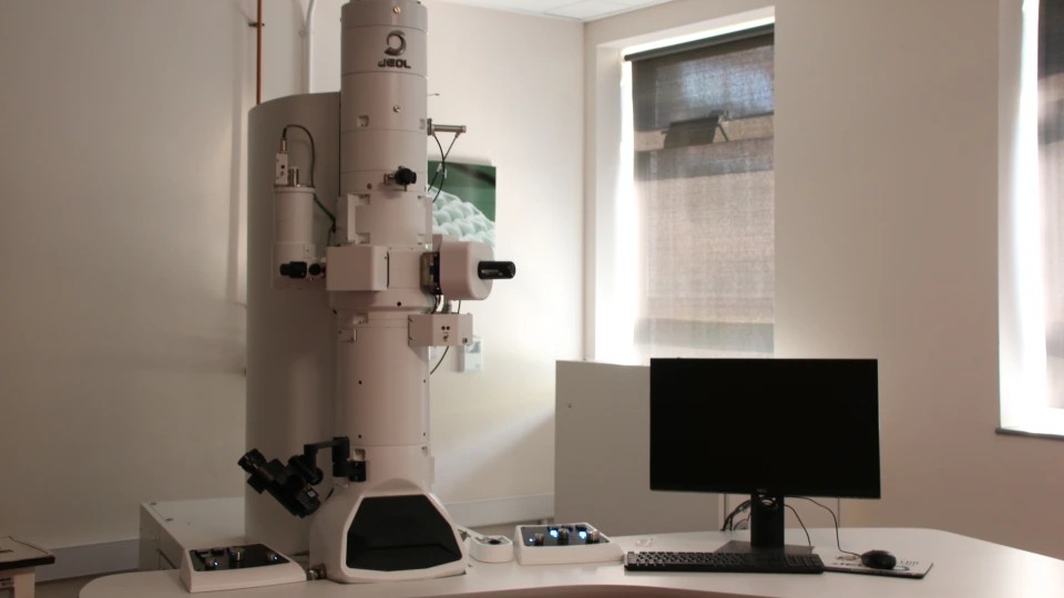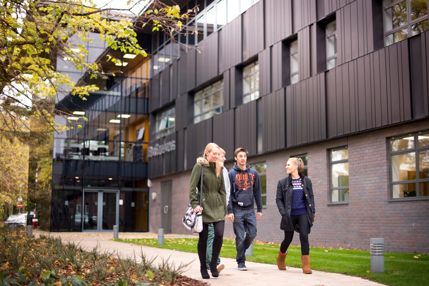
The imaging centre houses a TEM and two SEMs, facilities for on-site sample preparation and trained technical support. This centre is open to internal and external users from a range of backgrounds.
Interested in using the facilities? Please contact us.
What you can do here
The JEOL Imaging Centre supports researchers working across Biology, Chemistry, Geology and beyond. With capabilities in investigating ultra structures of biological tissue, characterising nanoparticles, or mapping the elemental composition of a material, our instruments and preparation facilities produce the resolution and analytical depth to help facilitate your research.
Our Equipment
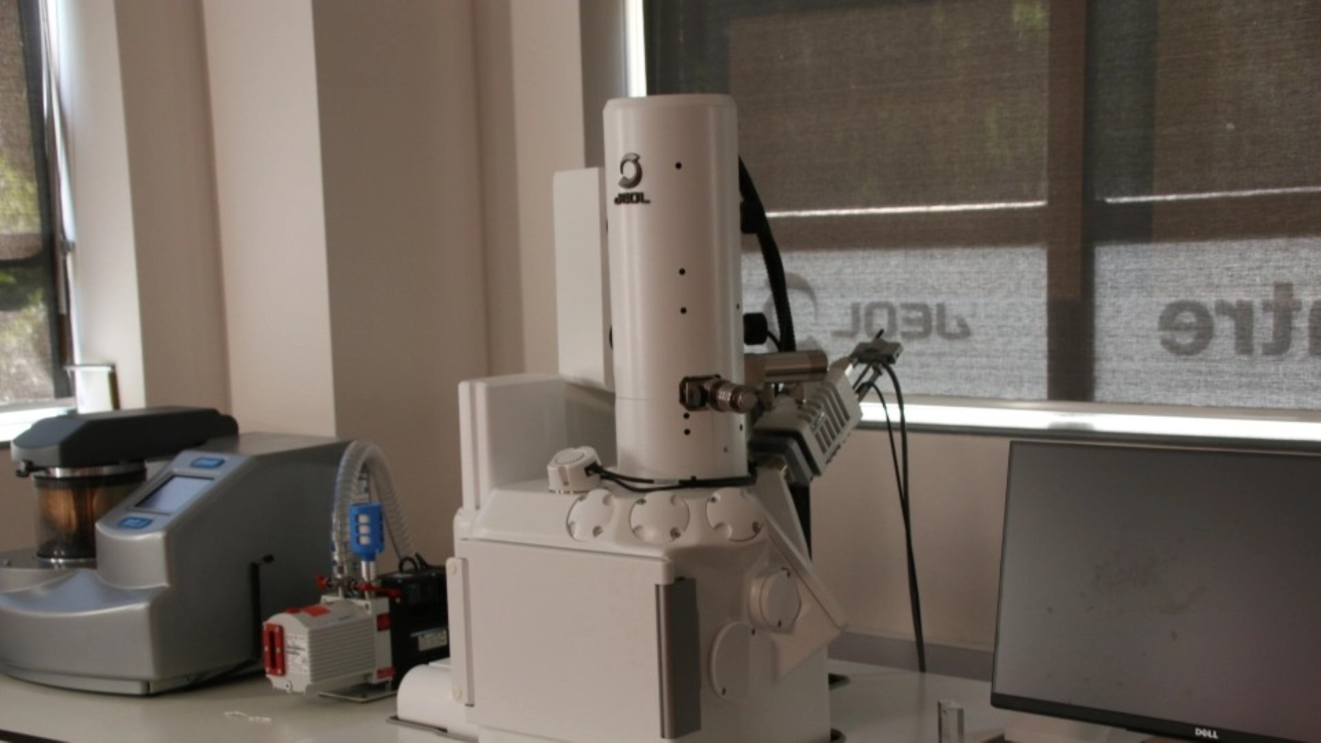
Our most advanced SEM, featuring a field emission gun system with integrated Energy Dispersive X-ray Spectroscopy (EDS) for real-time elemental mapping alongside high-resolution surface imaging.
Resolution: 1.0 nm Â
Magnification: up to 600,000× Voltage: 0.5–30 kV
Max specimen: 200 mm ⌀, 75 mm height
Low-vacuum mode: 1.8 nm
Real-time EDSÂ
An accessible tungsten filament SEM with a straightforward interface, this model is suited for users both new to electron microscopy, and experienced microscopists.Â
Resolution: 4.0 nm
Magnification: up to 300,000× Voltage: 0.5–20 kV
Max specimen: 152 mm ⌀, 49 mm height
Low-vacuum mode: 5.0 nmÂ
Our TEM delivers sub-nanometre resolution imaging of internal ultrastructure, ideal for biological sections prepared by ultramicrotomy, as well as nanoparticles and materials samples.
Resolution: 0.2 nm
Magnification: 10× to 1,200,000×
Voltage: 10–120 kVÂ
Sample Preperation
Successful electron microscopy depends on good sample preparation. The biology department offers dedicated equipment alongside the microscopes, and technical staff who can advise on the most appropriate workflow for your sample type.
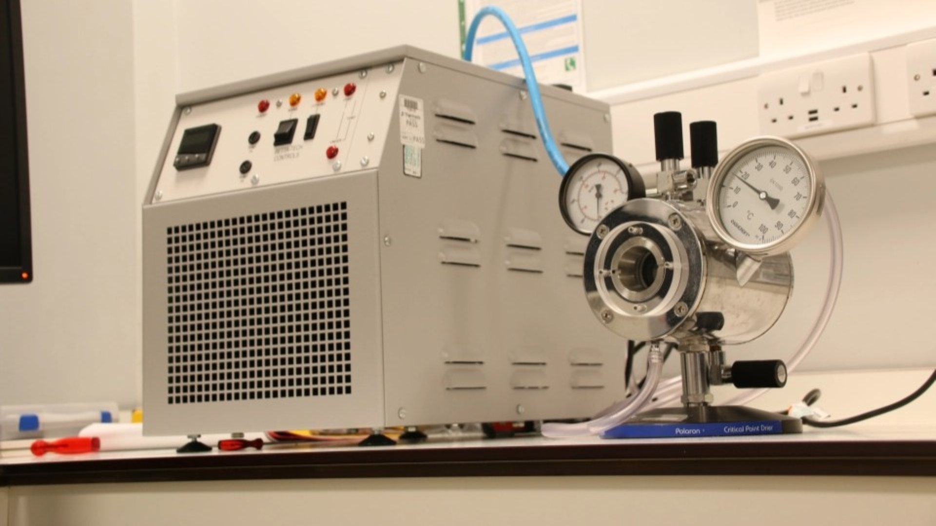
Currently available by prior arrangement – please contact us before planning use.
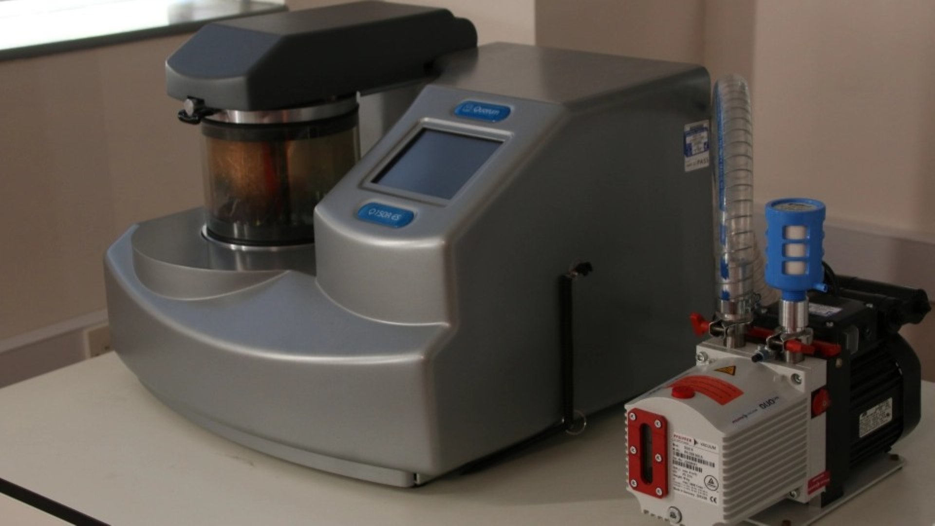
Quorum E300 Critical Point Dryer- Removes water from biological samples whilst preserving three-dimensional structure
-
Prevents surface tension artefacts, important for delicate tissues
- Applies a thin conductive metal coating (gold, gold/palladium) to non-conductive samples for SEM
- Also supports carbon coating for EDS compositional work
- Essential for biological and polymer SEM samplesÂ
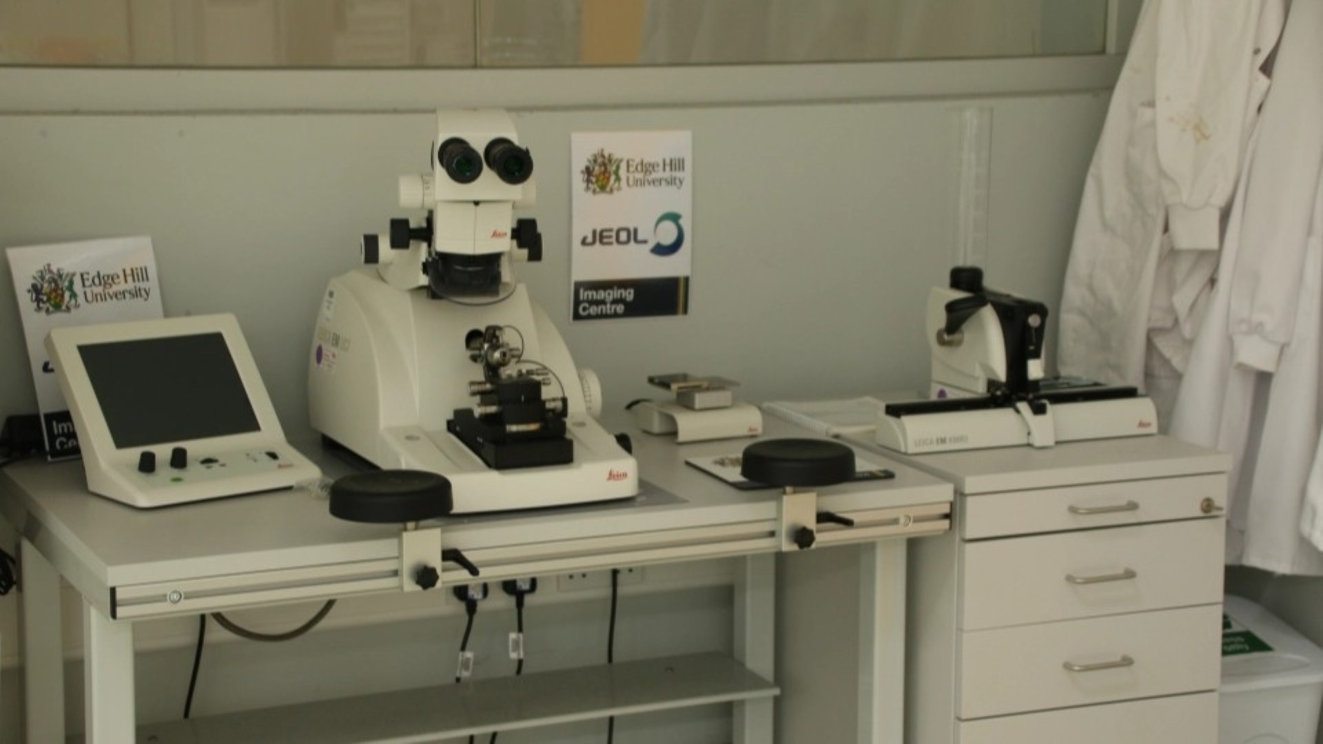
- Produces ultrathin sections (50–100 nm) from resin-embedded samples
- Essential for TEM imaging of cellular ultrastructure
- Suitable for biological tissue, polymers, and composite materialsÂ
Chemical Drying
Chemical alternatives to critical point drying for SEM sample preparation are available, and suitable for a wide range of biological samples. Please enquire for specific chemical drying protocols.