Histology Laboratory
A complete tissue preparation and analysis workflow from processing and embedding through to precision sectioning and brightfield imaging, all within a single dedicated laboratory in our Life Sciences Building.
What you can do here
Our Histology Laboratory supports the full tissue preparation pipeline; all centralised to our new Life Sciences building. Whether you’re working with mammalian tissue for biomedical research, ecological specimens, or novel materials requiring thin sectioning, our equipment and workflow cover every stage from fresh tissue through to stain sections ready for imaging.
Our equipment
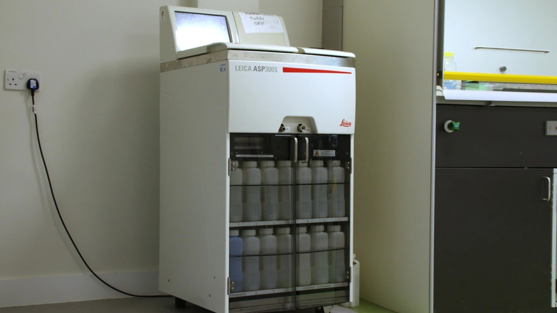
An automated closed-system tissue processor for fixation, dehydration, clearing, and paraffin wax infiltration of histological specimens. Programmable protocols allow overnight runs with consistent, reproducible results.
Suitable for a wide range of tissue types including soft tissue, bone, and plant material.Â
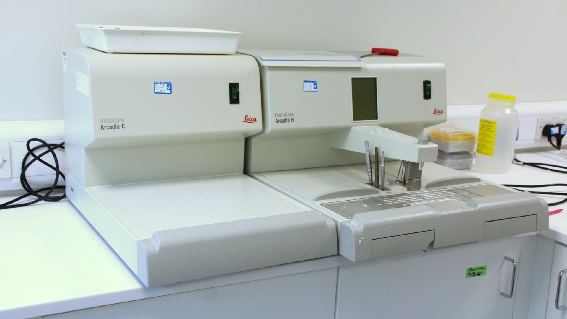
A microprocessor-controlled paraffin embedding station with heated forceps and a cold plate, allowing precise orientation of specimens within the mould prior to sectioning.
Produces blocks with consistent, flat faces, critical for achieving good section on a microtome.Â
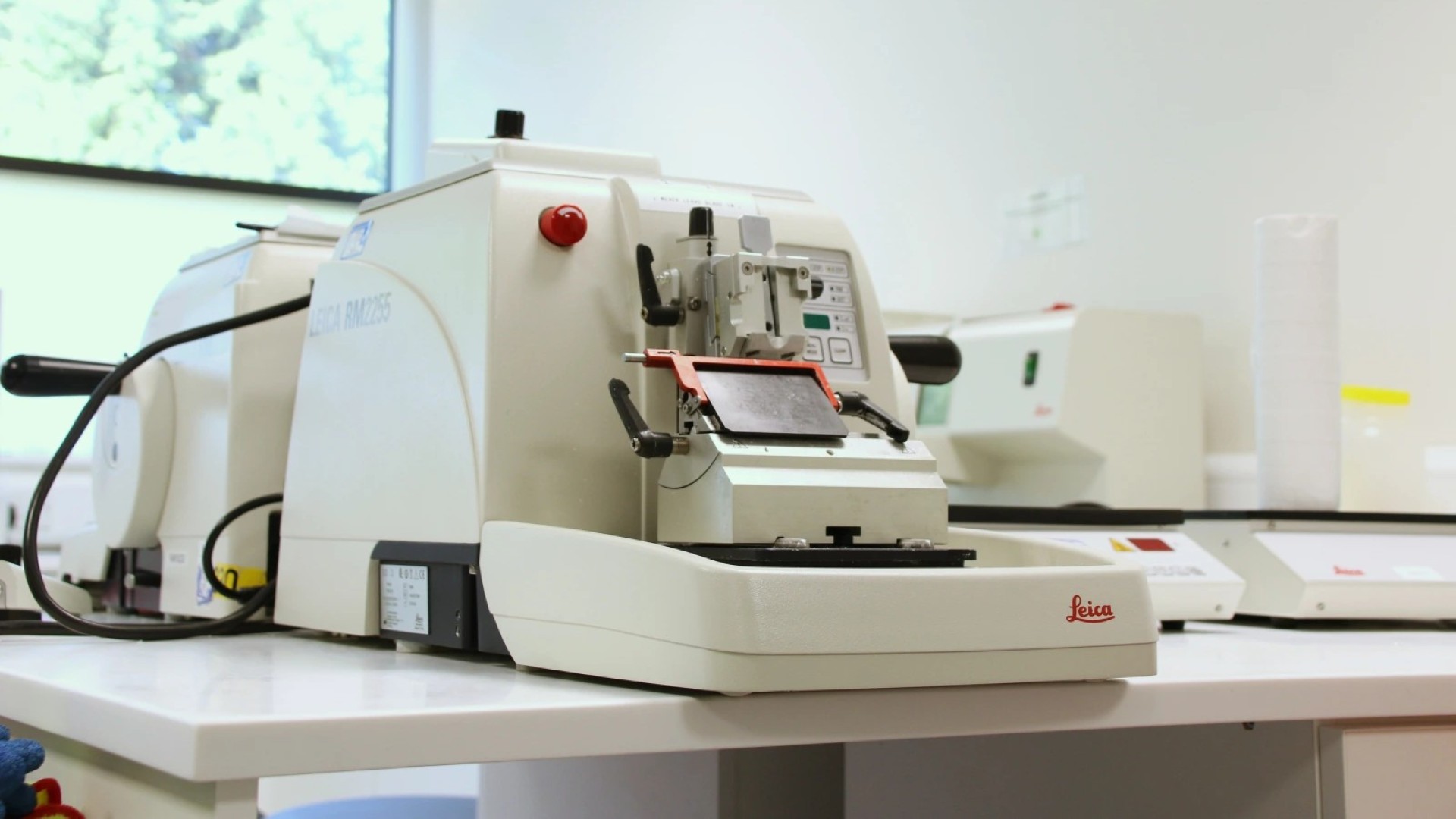
A fully automatic, motorised rotary microtome designed to produce thin, reproducible sections of paraffin-embedded specimens. Suitable for routine histological sectioning across a wide range of tissue types.
Section thickness range: 0.5–100 µm
Fully motorised advance
Programmable sectioning speed
Low-vibration cutting for consistent section quality
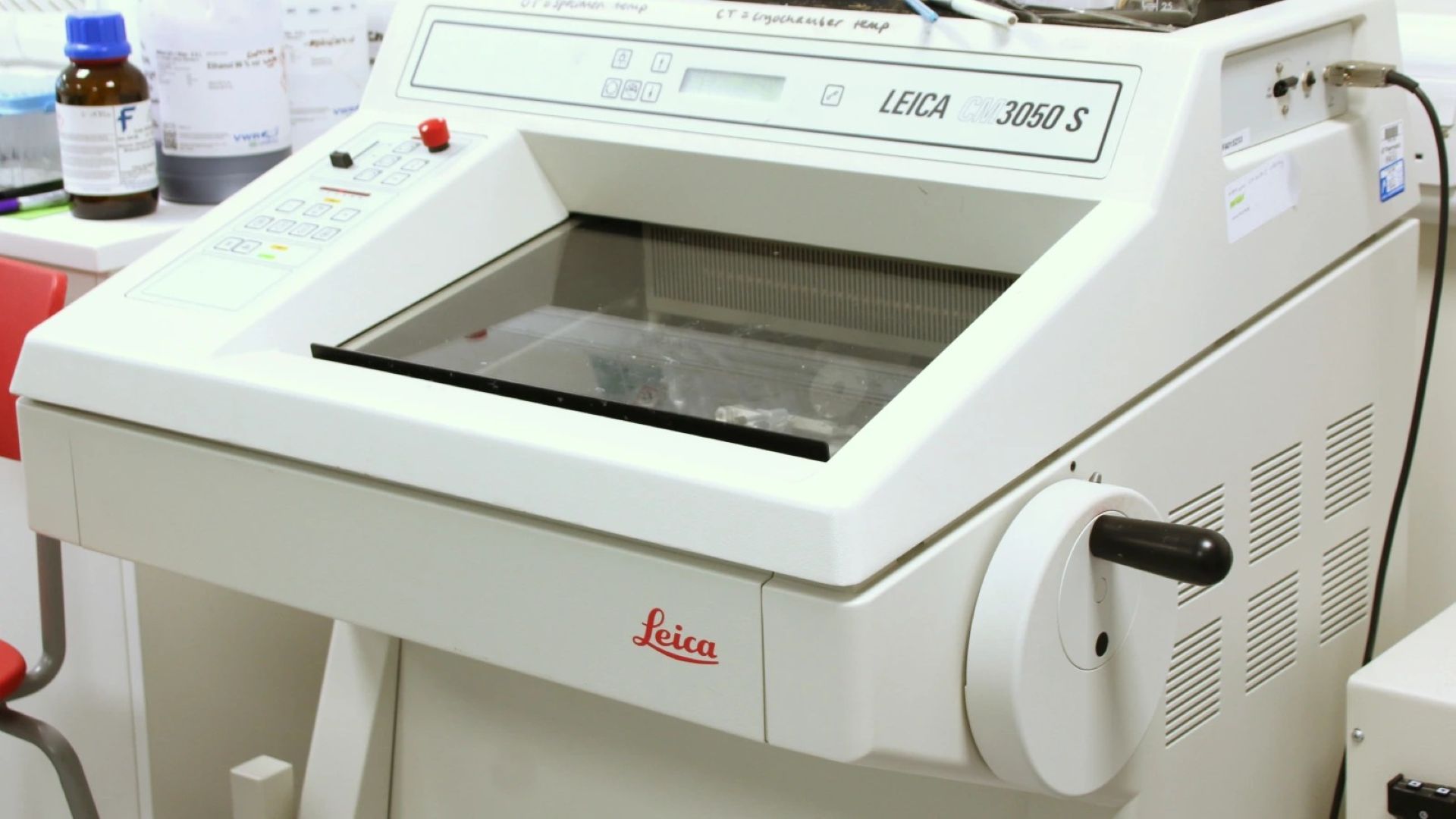
A motorised cryostat for rapid frozen sectioning, a preferred route where tissue processing time is limited, enzyme activity must be preserved, or antigen retrieval for immunohistochemistry is a priority. Â
Motorised drive Â
Section counter Â
x/y orientation ±8° Â
25 mm horizontal specimen feed Â
Independent specimen temp. control Â
Low-profile disposable blades