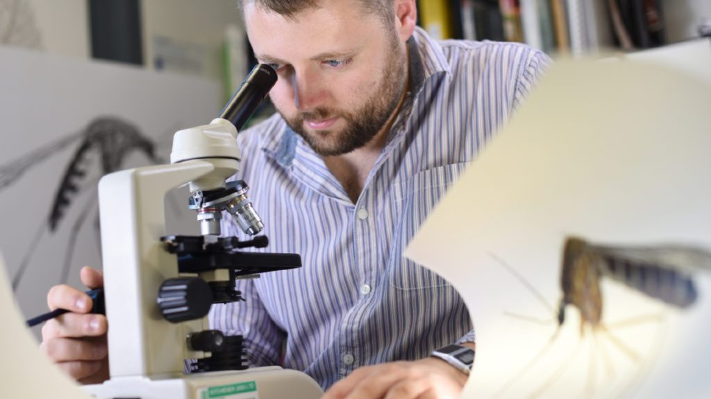
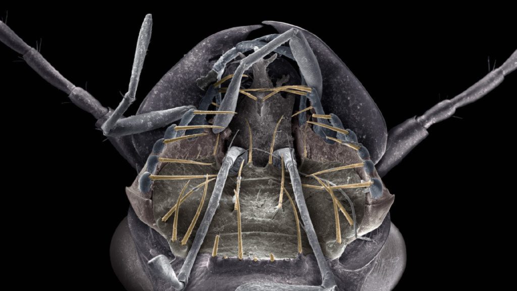
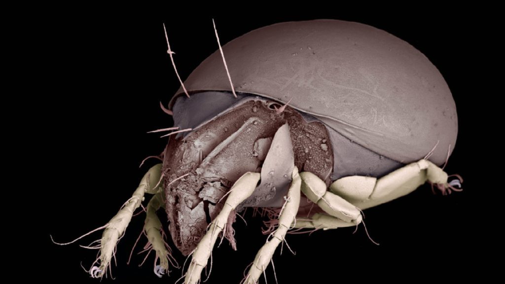
Dr Thom Dallimore is responsible for Fluorescence and Scanning Electron Microscopy within the department. He’s showing us around the Scanning Electron Microscope (SEM) facilities within the Department of Biology. He points to pictures scattered around the walls: there’s the mouth of a small beetle; that’s a mite; here’s a mosquito.
All fractions of centimetres in reality, blown up in hugely impressive detail. To give you an idea of the power of these things, a traditional microscope which uses light, you know, the kind you find in school labs, will give you something like 1000x magnification. These will give you something like 100,000x magnification. Mind blown.
“It makes you feel a bit insignificant. You know when people try and think about the concept of the universe and how vast it is, it makes people feel dejected and isolated and totally unimportant. Here you’re looking at things that are so small and you find these worlds, these microcosms, that are unbelievably tiny, and you feel equally insignificant.”
Dr Thom Dallimore
And now the University is about to go to the next level, in partnership with JEOL Imaging Centre, the high-powered microscope manufacturer. With the arrival of two brand new next-gen microscopes – another, more sophisticated SEM and a transmission electron microscope (TEM) – we continue to develop our state-of-the-art facilities. Both microscopes come in at a cool £150k each, with the new SEM the first of its kind in the country.
A molecular biologist at heart, Thom “just mucks about making lovely pictures”, but of course these microscopes are here to add another dimension to the learning experience of our students, as well as improving the department’s research capabilities.
We run lab masterclasses for our undergraduate students. They’re taught how to use high-powered microscopes. And that was a reason we were keen to get another one. They all collect something that they think is interesting – a fly, or their own hair, or a grain of rice.
So what can these microscopes do that they other ones couldn’t?
The new SEM has a chamber about twice the size. Pizza oven size. That’s really useful. Normally you have to go through a fairly tedious process of sample preparation, mostly to remove moisture, because when you fire electrons into something that’s wet, you get this image that just glows white, and you can’t see anything. At the same time, you don’t want it to shrivel up, so we have these chemical ways of removing moisture without destroying the structure. They’ve recently developed this new system of drying samples by freezing them in liquid nitrogen, a bit like the T-1000 in the Terminator films, and put them straight into the imaging chamber. No chemicals. And that’s revolutionised high-powered microscopy because it means you can look at things that are wet. So in the long run we’d like to get this liquid nitrogen tech in here as well, and the bigger chamber allows us to do that.
You had us at T1000, Thom…
The transmission electron microscope fires electrons into the surface of a sample and slices super thin slivers of material. For example, if you wanted to look at the cells in a human ear, you’d take a chunk of ear, embed it in a resin, and then the machine fires electrons through superfine slices. Not only can you see the structures within cells, but you can see the organelle [a functional unit in a cell or unicellular organism, such as a mitochondrion] details, the DNA structures, all that kind of stuff. So if you want to look at how cells work, it’s perfect.
Like the T-1000, though, the new microscopes have more tricks up their sleeves. They come equipped with energy-dispersive X-ray spectroscopy capability (or EDS, to you and I). This enables you to map the chemical composition of whatever you’re looking at, says Thom:
If you’ve got a rock and want to know what it’s made of, then it’ll identify the different chemicals and minerals. And in biology that can be really useful. If you’ve got a tiny beetle that produces a pheromone, and you want to know where it’s produced and what it’s made of, you can image it, and see where the hotspots of this particular chemical are. It’s pretty impressive.
And there are a multitude of other things to examine through a SEM/TEM.
All undergraduates are able to take the Laboratory Masterclass module. Megan Quail, now an MRes student, used the SEM to study the structural differences between the wool fibres of various sheep breeds, something that hadn’t yet been explored to any great depth.
And Plant Science students will be looking at plant cells, which is a completely different field, continues Thom:
These microscopes have revolutionised biology. We can see structures in cells we never even knew were there. And we can carry out experiments to determine what they do, what their functions are. Anything to do with cells, it’s fantastic.
And other disciplines within the University have expressed interest in getting some quality time with the new machines. The Geography and Geology department can use it to examine mineral structures in rocks. The new Engineering team could be using it to examine the structures of materials, and the Medical School may actually be checking out ear cells, among other things.
And there are commercial interests. Those working in the medical and engineering industries will be using our microscopes to find out how effective the anti-microbial surfaces on medical tubing are, or why their calculations, based on maths, physics and chemistry, aren’t quite working out. A food manufacturer has used the microscopes to discover why their ice cream was developing a grainy texture in the transition from factory to shop: “Troubleshooting, quality control – they’re really useful for those kind of things,” says Thom
So, the million dollar question, Thom – well, £150,000 pound question, at least: are you not worried about letting your undergraduates loose on these machines? Thom’s very clear that these are very much working machines.
They’re surprisingly robust. When something goes wrong, it really goes wrong. We don’t play the blame game, though – accidents are going to happen. If you want people to use it, you have to be prepared for it.
And with that Thom is back to making what he modestly refers to as “nice pictures”:
For a scientist this is like going on holiday to Legoland [other holiday parks are available]. You can see structures that you’ve never seen before, and it just blows your mind. Like those mites, those things are like full stops, and even under a light microscope they still look like full stops, but under here, the level of detail just brings everything to life.
June 10, 2022