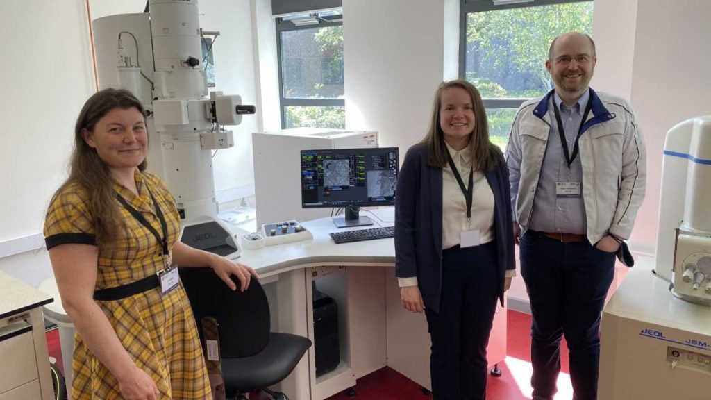
Following a £500,000 investment, the University has purchased two brand new next-gen electron microscopes – a transmission electron microscope (TEM) and a scanning electron microscope (SEM), the first of its kind in the country. They have been manufactured by JEOL, a world leader in electron microscopes and analytical instruments.
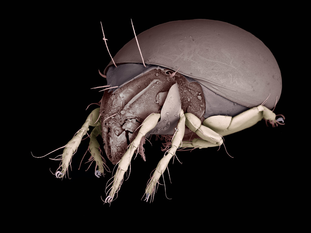
The electron microscopes allow Edge Hill’s scientists and students to take detailed images at over 100000x magnification – much more advanced than the 1000x magnification a traditional microscope allows for. The images can be used to learn more about the microscopic organisms and objects that surround us.
The new facilities will be used by JEOL as a specialist training centre, providing a unique opportunity for students to get hands-on experience with industry-standard equipment and work more closely with employers in the sector to enhance their employability.

Professor Paul Ashton, Head of Biology, said: “It’s been 10 years since Biology became an independent department and it’s amazing to see how far we’ve come. We’ve attracted significant financial investment and the hundreds of biology students we teach will now have access to a state-of-the-art imaging centre with direct links to industry leaders JEOL.
“This will support us in our mission to give biosciences students the best possible experience. The equipment will also enhance our biomedical research and open up new areas of investigation to our academic community.”
Within the Biology department, the electron microscopy equipment will be used for imaging the surface of cells and nanomaterials. This type of microscopy is used for research in fields such as regenerative medicine, drug development and nanomedicine.
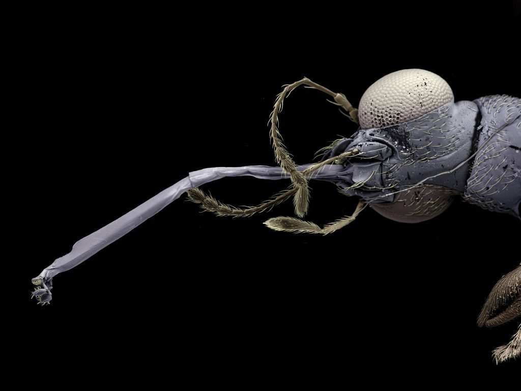
A team of academics from Edge Hill used the existing SEM to conduct research into invasive Aedes mosquitos, which are vectors for spreading major diseases. With it, the researchers were able to identify microscopic eggs on old tyres which were being traded all over the world. The research led to advancements in finding and destroying mosquito eggs on a huge range of surfaces and products.
The TEM has uses far beyond biosciences, supporting research in Engineering and Medicine.
To discover more about our courses at Edge Hill, please visit ehu.ac.uk/study.
May 12, 2022